← Back to home

Percutaneous techniques

Minimally invasive foot surgery

Correcting selected deformities through small incisions, using dedicated instruments and imaging guidance — when this approach is appropriate for the condition and the patient.

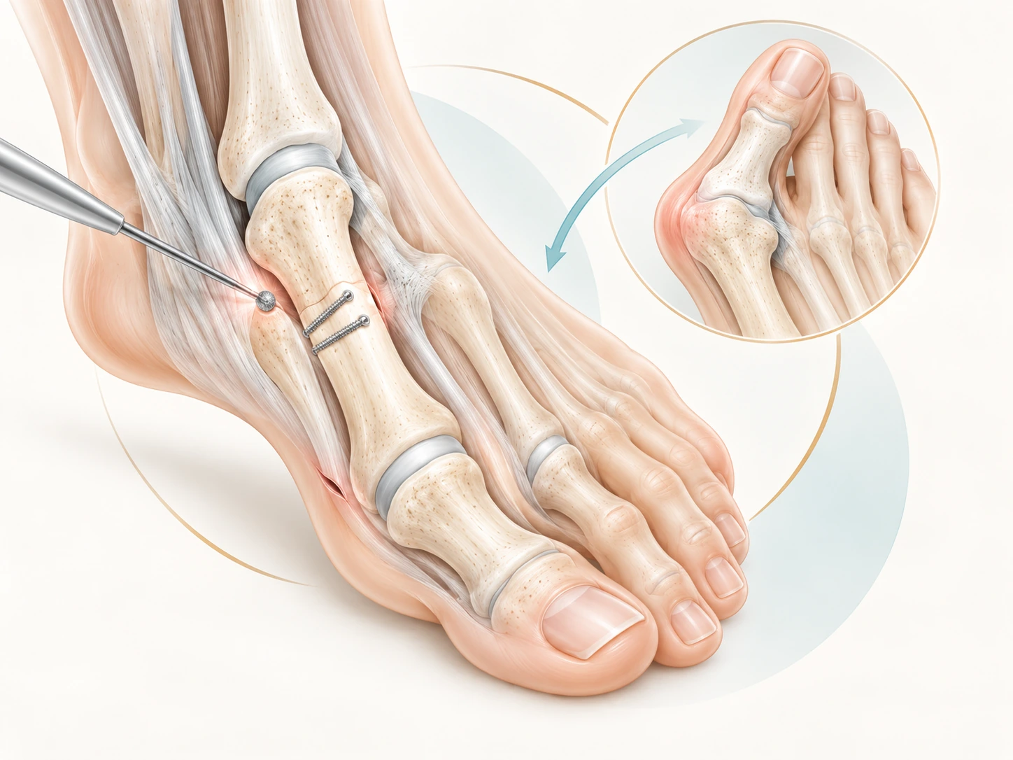
Educational illustration of percutaneous hallux valgus surgery
Principle

What is minimally invasive surgery?

Minimally invasive surgery (MIS) uses small incisions to perform bone or soft-tissue procedures with dedicated instruments. In percutaneous surgery, some steps are performed without widely exposing the bone.

The surgeon relies on three-dimensional anatomical knowledge and, depending on the procedure, fluoroscopy to check correction and implant position. 'Minimally invasive' therefore describes the surgical approach: it does not mean minor surgery, no risk, or instant recovery.

The key principleChoose the technique that achieves the appropriate correction with the least necessary tissue disruption — not a small incision at any cost.
Indications

When can it be used?

The indication depends on the deformity, its severity, bone quality, the joints involved and the patient's goals.

Hallux valgus

Percutaneous osteotomies of the first metatarsal and, when indicated, the proximal phalanx.

Metatarsalgia & lesser toes

Selected metatarsal osteotomies and lesser-toe deformity corrections.

Hindfoot

Selected calcaneal osteotomies can be performed through minimally invasive approaches.

Other procedures

Selected fusions or targeted procedures may benefit from a minimally invasive approach, depending on anatomy and surgical expertise.

Benefits

What benefits can be expected?

Less exposure

Smaller incisions and limited soft-tissue dissection.

Early pain

For hallux valgus, some evidence suggests less pain in the early postoperative phase.

Same objective

Achieve a durable, functional correction suited to the anatomy — just as with open surgery.

A smaller scar does not mean recovery without swelling or restrictions. Bone still needs to heal after an osteotomy.

Surgery

What happens during surgery?

Planning

Clinical examination and weight-bearing radiographs to understand the deformity as a whole.

Small approaches

Instruments are introduced through several millimetric or short incisions, depending on the procedure.

Bone correction

A dedicated burr may be used to perform the osteotomy. The bone is translated into the planned position.

Fixation & verification

Correction is usually stabilised with screws and checked radiographically before closure.

Clinical example

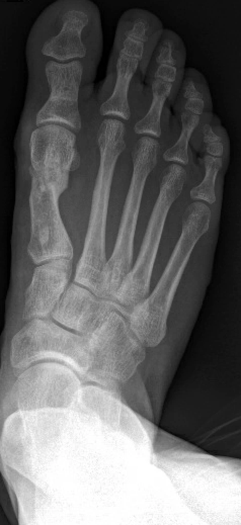
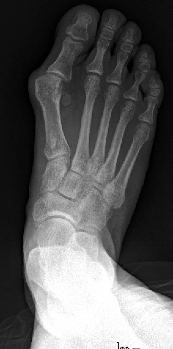
Hallux valgus: example before and after surgery

Right-foot radiographs illustrating a preoperative and postoperative example. Results and technique are individualised according to the deformity and the patient.

Preoperative X-ray of right-foot hallux valgus
Before surgery
Postoperative X-ray of the right foot after hallux valgus correction
After surgery
Recovery

Recovery depends on the procedure performed

After hallux valgus correction, protected weight-bearing in a postoperative shoe can often begin early. The exact protocol depends on the osteotomy, fixation and additional procedures.

0–2 wksProtect

Elevation, swelling control, wound care and weight-bearing as prescribed.

2–6 wksHeal

Progress guided by wound healing and radiographic review.

6–12 wksResume

Transition towards normal footwear and gradual return to activity according to progress.

Several monthsSettle

Foot swelling can persist for several months even when bone healing is satisfactory.

Risks

Limitations and risks

Possible complications include infection, nerve irritation or injury, delayed or non-union, displacement or loss of correction, stiffness, persistent pain, recurrence, hardware-related problems and further surgery. Some risks are specific to the procedure performed.

Patient selectionMinimally invasive surgery is an additional option in the surgeon's armamentarium, not a universal replacement for open surgery. Some deformities are better treated through an open or combined approach.

Selected medical sources

  1. Tham A et al. Minimally invasive chevron osteotomy provides comparable outcomes to open surgery for hallux valgus: systematic review and meta-analysis. Foot Ankle Surg. 2026.
  2. Zverev SR et al. Outcomes of minimally invasive versus open hallux valgus surgical correction: systematic review and fragility analysis. J Foot Ankle Surg. 2026.
  3. Lewis T et al. Minimally invasive percutaneous surgery techniques for common foot & ankle pathologies: Current concepts and clinical outcomes. 2026.
  4. Cardoso DV et al. Cohort comparison of radiographic correction and complications between minimally invasive and open Lapidus procedures for hallux valgus. Foot & Ankle International. 2022.

Helping you move forward, step by step.

A consultation is an opportunity to discuss your situation, goals and the different treatment options.

Specialist consultation

Which technique for your foot?

The choice between minimally invasive, open, or combined surgery follows clinical examination and weight-bearing imaging.